LAPAROSCOPIC HERNIA REPAIR
What is an incisional/ventral hernia?
Incisional/ventral hernias are defects that appear at the site of a prior surgical incision.
An incisional hernia can develop in the abdominal wall around a previous incision. It usually arises in the abdominal wall where a previous surgical incision was made. This results in a bulge or a tear in the area where the abdominal muscles have weakened. Incisional hernias can increase in size with time.
A hernia occurs when part of an organ (usually the bowel or intestine) protrudes through a weak point or tear in the thin muscular wall that holds the abdominal organs in place.
Hernias can develop around the navel, in the groin, or any place where you may have had a surgical incision. Some hernias are present at birth. Others develop slowly over a period of months or years. Hernias also can come on quite suddenly.
What are the symptoms of a hernia?
An incisional hernia causes a bulge in the abdominal area. Often, this type of hernia is painless, but may be tender and can cause discomfort during any type of physical strain, such as lifting heavy objects, coughing, or straining during bowel movements.
The bulge may disappear when lying down, and be more visible when standing up.
Symptoms include pain, which may be a sharp or dull ache that feels worse towards the end of the day. Other symptoms include nausea, vomiting, and the inability to have a bowel movement. Continuous or severe discomfort, or nausea related to the bulge are signs that the hernia may be entrapped or strangulated. If you have these symptoms, be sure to contact your physician immediately.
How is a laparoscopic ventral hernia repair performed?
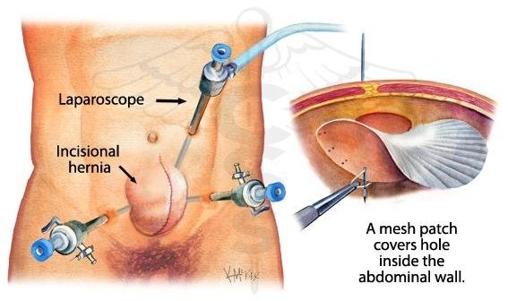
A surgeon uses special instruments, small incisions, and a videoscope and television monitors to perform the hernia repair. A small incision is made in the abdominal wall in a location chosen to minimize the risk of running into organs or scar tissue from prior operations. Surgeons make as few other tiny incisions as is feasible, depending on how much scar tissue there is and how well they can see.
A laparoscope (a tiny telescope with a television camera attached) is inserted through a small hollow tube. The laparoscope and TV camera allow the surgeon to view the hernia from the inside.
Other small incisions will be made for placement of other instruments to remove any scar tissue, and to insert a surgical mesh into the abdomen. This mesh is fixed under the hernia defect to the strong tissues of the abdominal wall. The surgeon will use mesh to reinforce the weakened area of the abdominal wall. This will help prevent the hernia from recurring.
What are the advantages of the laparascopic approach?
There are many advantages to this approach, including quicker recovery and shorter hospital stays, as well as a significantly reduced risk of infection and recurrence.
Patients usually go home within 24 hours after laparoscopic repair, as opposed to a longer hospital stay after open repair, and report less pain and quicker return to normal activity.
What is the chance of recurrence, compared with the traditional approach?
The rate of recurrence is much less with the laparasocpic approach (less than 10%) as compared to the 20-40% recurrence rate with the open procedure.
Related Services
-
LASER PROSTATECTOMY: THE “BLOODLESS” PROSTATE SURGERY
A prostatectomy is the surgical removal of all or part of the prostate gland. Abnormalities of the prostate, such as a tumour, or if the gland itself becomes
-
Minimal Access Advanced Laparoscopic Surgery
Fibroids are a common uterine tumor affecting 20 to 25% women. Fibroid develops from benign transformation of a single smooth muscle cell. The growth of Myoma is dependent on many factors. Increased estrogen stimulation alone or together with growth hormone or human placental lactogen appears to be the major growth regulator of Fibroid.
