
Fetal Diagnostic And Ultrasound Services
Gynaecologic ultrasound scan
The gynaecologic ultrasound scan is carried out to examine the organs of reproduction in the woman who is not pregnant. This examination looks at those pelvic organs like the cervix, uterus, tubes and the ovaries and detects normalcy or otherwise in these structures. Abnormalities like ovarian cysts, ectopic pregnancy, uterine fibroid and suspected ovarian cancers are some of the diagnosis that can be made with this ultrasound scan.
The gynaecologic scan usually is done first with a transabdominal scan which helps to get a good view of the whole of the pelvis. In many instances, the transabdominal examination is followed by a transvaginal examination. This involves using a slender probe that is introduced into the vagina to view the pelvic structures. This examination requires an empty bladder hence women are not subjected to drinking water and the delay that it necessitates. With this combination of approaches, most gynaecologic diagnoses can be made accurately.
Uterine anomalies: A gynecologic ultrasound scan can diagnose some congenital (inborn) abnormalities of the womb. It could also help diagnose women with no menses even after puberty who may be retaining the menses (like in imperforate hymen).
Uterine fibroids: constitute the bulk of uterine abnormalities in our environment. Ultrasound can help diagnose the presence of the fibroid, its size and location, presence of complications and if it is likely to interfere with pregnancy or delivery.
Infertility work up: A gynaecologic ultrasound scan is important in the evaluation of a woman who is being managed for infertility. The uterus and other pelvic organs are assessed for abnormalities. In addition, a transvaginal scan can be used in follicular tracking to know if the woman is likely to be ovulating. This may guide the treatment offered.
Hystero-contrast sonography: This technique uses ultrasound scan to assess the cervix (mouth of the womb), uterine cavity (inside of the womb) and the tubes. This is very important in women being evaluated for infertility. This test could be used as an alternative to the traditional hysterosalpingography (HSG) which is loathed by many women because of the pain it causes. This test can also accompany a HSG if the findings are ambiguous. It may also obviate the need for a laparoscopy and a dye test which is an operative procedure.
Cardio-tocography (CTG)
This is a special type of examination that studies the pattern of the fetal heart rate. The essence of the examination is to determine the health of the baby at a particular point in time. It can also predict how much time one can delay within which the baby is unlikely to die except an emergency like bleeding occurs. It consists of a probe that is attached to the abdomen and held in place with a belt. It continuously monitors the heart rate of the baby over a period of time. Specific patterns are expected of a healthy baby. Deviations from this pattern can be recognized and action taken. For high risk pregnancies, the CTG can be repeated frequently sometimes weekly and at other times, it can be repeated daily depending on the risk.
DETERMINATION OF SICKLE CELL STATUS OF THE BABY
It is well known that the surest way to avoid having a baby with sickle cell disease is for an AS person not to marry another AS or SS individual. However, for certain reasons, some persons still go ahead with marriage or are already married. Since the offspring of this union has a chance of having sickle cell disease, the parents may want to know before the baby is born. The earliest time the tests can be done in pregnancy is about 12 weeks. A CVS (earlier discussed) is taken and the samples sent for a special test to determine if the sickle cell gene is present. This information is highly valued by many parents who will like to know early what to expect and start preparing their minds for it. However, for many, it makes no difference. In this latter group, doing such test is not necessary.
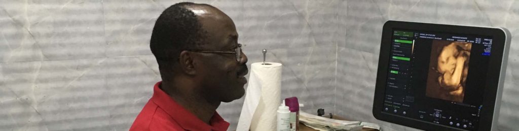
3D and 4D scans
The 3D and 4D scans make use of more advanced ultrasound technology. It enables us to see the various structures more clearly giving it a 3 dimensional view as if being seen in real life. It is very exciting to mothers to see a live 3 dimensional view of their babies especially the face. This added social benefit is of value to a lot of mothers.
This center does not offer a 3D or 4D service for now. This is partly because the 3D/4D scans increase significantly the cost of ultrasound and also because of what we consider as the limited medical related information that it will offer in our setting at present. It gives added medical benefit after a thorough 2 dimensional scan especially if there are structures that require more characterization. However, as the uptake of advanced ultrasound improves, there will be need in the future to upgrade to the 3D/4D scans.
Storage of images
Some of the essential images generated during the scanning of a client are stored electronically for a period. This may become useful in future if there is need to review the images at a later date.
Generation of reports
Generation of reports is an essential aspect of the diagnostic procedure. A good report helps to identify the major issues in a scan and convey such to the managing doctor. We use the astraia software [astraia medical software, Germany] for the generation of our reports. The software highlights areas of concern, provides charts and centile values that make comprehension of growth pattern of the fetus obvious and gives a comprehensive overview of the scan findings.
More Services
-
Bloodless Medicine And Surgery
Safehands Medical Centre has extensive and proven experience in patient blood management and is actively involved in the provision of bloodless medicine and surgery(BMS)
-
Anesthesiology
Anesthesia means lack of sensation. It prevents patients from feeling the surgery or medical procedure. Normally, nerves from every part of the body