
3D / 4D Ultrasound and Pregnancy
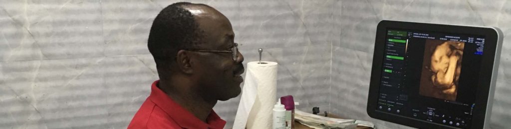
3D / 4D imaging is a relatively recent development in ultrasound technology. In 3D ultrasound imaging, a series of multiple slices of 2D slices are taken and stored digitally. These can then produce life like pictures. 4D scanning simply means that theses life like pictures can be seen to move in real time and hence the activity of the baby can be studied.
Is 3D / 4D scanning safe in pregnancy?
According to the available literature, there are no adverse effects on the fetus from using this technology as the same type and intensity of ultrasound is used as with conventional 2D imaging. In fact, 3D scanning should reduce the exposure time as it allows storage of the data for later analysis without continual scanning.
What is the best time to have 3D / 4D scan?
The optimal time is considered to be 22- 26 weeks as the amniotic fluid volume is optimal and baby tend to have more subcutaneous fat.
What are benefits of 3D / 4D scanning?
The benefits of 3D / 4D scanning in obstetrics are still not well defined unlike in Gynaecology.
 |
 |
 |
Some of the benefits are:
- Face, arms, fingers, legs and toes are seen more clearly
- Activities such as moving limbs, yawning, swallowing can be seen with 4D scanning.
- Bonding between parents and baby has shown to be stronger because of more realistic picture of the baby.
Will 3D image always be better than conventional scan pictures?
Though the 3D imaging is attempted at all scans, it can become extremely difficult in late third trimester. The factors that affect the quality of the images are fetal position, amount of fluid, placental position and if you are overweight.
We are a medical specialist ultrasound practice and we offer 3D / 4D ultrasound only as part of an ultrasound examination for clinical reasons requested by a medical practitioner.
Related Services
-
Cervical Length Assessment in Pregnancy
Preterm birth complicates about 8 to 10 percent of all births. A short cervix (< 25mm) or dilated cervix early in the second trimester of pregnancy appears to be a warning sign of impending premature birth among women who have previously given birth prematurely.
-
Early Pregnancy Scan / Dating Scan
Early Pregnancy Scan / Dating Scan is a scan done early in pregnancy before 12 weeks of gestation. It is otherwise known as a dating/ viability scan.