
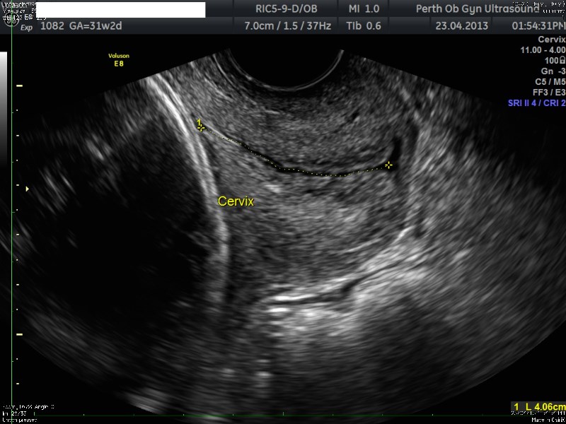
Cervical Length Assessment in Pregnancy
Preterm birth complicates about 8 to 10 percent of all births. A short cervix (< 25mm) or dilated cervix early in the second trimester of pregnancy appears to be a warning sign of impending premature birth among women who have previously given birth prematurely. Cervical length assessed by transvaginal sonography between 14 weeks and 24weeks, augmented by serial evaluations, predicts spontaneous preterm birth before 35 weeks’ gestation in high-risk women. The rate of preterm delivery at < 34 weeks’ gestation increases dramatically when the cervical length is ≤ 1.5 cm.
When is cervical screening indicated during pregnancy?
- Previous preterm births < 34weeks
- Women with suspected cervical incompetence
- Women with cervical suture
- History of previous cervical surgery (Eg. Cone biopsy)
- Multiple pregnancies
What is the normal cervical length during pregnancy?
It varies with the gestational age
At 20 weeks the average length is 40mm
At 34 weeks the average length is 34mm
What is the cut off limit for short cervix?
When the cervical length is < 25mm, it is considered as a short cervix.
It is very abnormal when the length is < 15mm when the gestational age is < 24weeks.
What exactly is the information obtained during cervical assessment?
- Length of the cervix
- Dilatation of the cervix
- Funneling of membranes into the cervical canal
- Location of cervical suture if it is in place already
What preparations are necessary for the scan?
A transvaginal scan is mandatory to accurately assess the features of the cervix as described above. The bladder is emptied prior to the scan.
What happens if I am found have a short cervix?
We would inform your obstetrician about the findings as soon as the scan is done. Your Obstetrician may consider hospitalization, bed rest, cervical suture, medications, modifications of physical activity depending on your individual circumstances.
Related Services
-
Fetal Diagnostic And Ultrasound Services
The gynaecologic ultrasound scan is carried out to examine the organs of reproduction in the woman who is not pregnant. This examination looks
-
Placental Localisation and Low-Lying Placenta
A low-lying placenta is often diagnosed on ultrasound scan before 20 weeks. As the baby grows the placenta is carried upwards. For some women the placenta continues to be in the lower part of the womb in later gestation.